

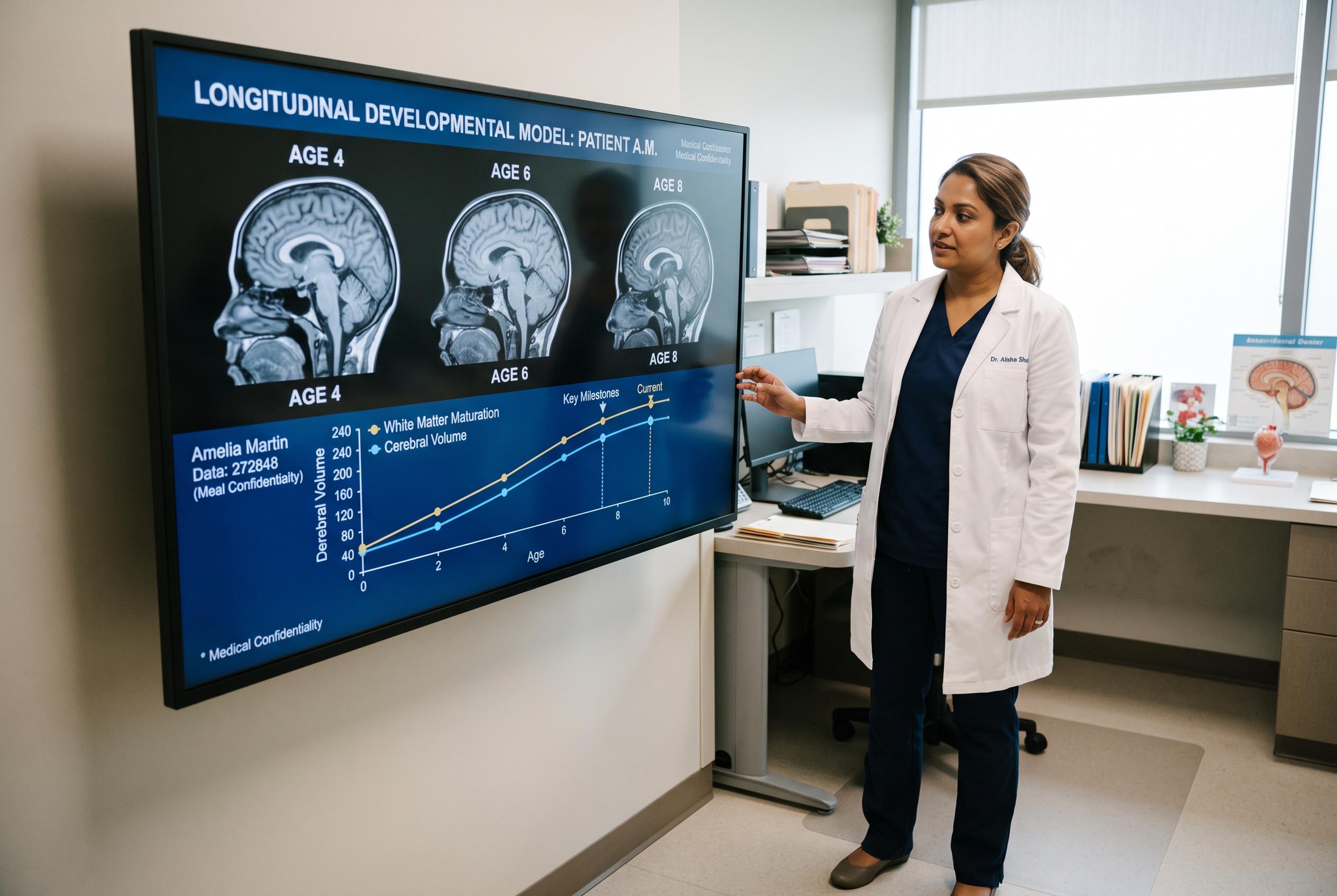

Researchers from the University of Pennsylvania unveil new MRI-based models that can predict atypical brain development in children years before clinical signs appear, opening new avenues for early intervention.
Researchers at the University of Pennsylvania say MRI-based longitudinal models may offer a way to spot children whose brain development is drifting away from expected growth patterns, potentially years before those differences become clinically obvious.
The work, published on August 7 in JAMA Network Open, builds on the idea that brain maturation is not a single, fixed path. Instead, the team led by Eren Kafadar and Aaron Alexander-Bloch used repeated MRI scans from the Adolescent Brain Cognitive Development study to compare each child’s changing brain volumes with normative growth curves derived from a large paediatric sample. The researchers said this conditional-longitudinal approach can capture individual departures from typical development more effectively than a single scan can.
In the study, baseline imaging from 10,830 children and follow-up scans from 7,262 participants were analysed alongside birth data and measures of psychopathology. The findings showed that lower birth weight was associated with lower longitudinal percentiles, suggesting greater-than-expected reductions in brain volume over time in 27 regions. Lower longitudinal percentiles were also linked with worsening psychiatric symptoms in 37 regions. By contrast, cross-sectional measures did not show the same associations, underscoring the value of tracking change over time rather than relying on one-time comparisons.
The result fits with a broader body of paediatric neuroimaging research showing that brain development varies substantially between children. A PubMed-linked analysis of ABCD data has highlighted the importance of accounting for age, sex and puberty when studying early adolescence, while other studies have used MRI to identify age-related differences in autistic children, track microstructural brain changes and estimate “brain age” as a marker of maturation. A Nature study spanning multiple cohorts has also linked infant and early childhood brain structure with cognition and adverse birth outcomes, reinforcing the view that development is shaped by both biology and early-life risk factors.
The Penn team said its data and code are publicly available and argued that the same framework could be applied beyond childhood development, including in studies of ageing. They suggested the method may help researchers ask not only whether a child’s brain looks different, but whether it is changing differently over time , a distinction that could matter in psychiatry, neurology and future screening efforts.
Disclaimer: This content is for informational purposes only and is not intended to be a substitute for professional medical judgment, advice, diagnosis, or treatment.