

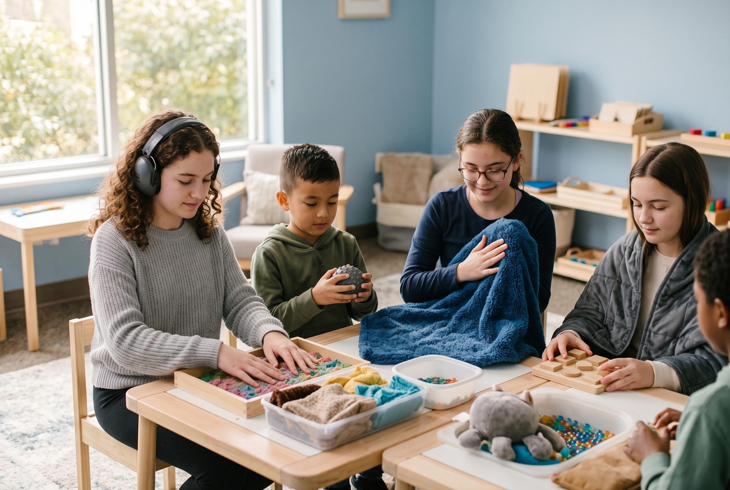

A study of 146 autistic children and teenagers found that distinct patterns of brain-network connectivity could predict differences in sensory experiences.
A recent study suggests that the very different sensory experiences reported by autistic children and teenagers may be tied to distinct patterns of brain connectivity. Writing in Biological Psychiatry: Cognitive Neuroscience and Neuroimaging, researchers reported that functional links between cortical and subcortical regions were strong enough to predict which sensory profile an autistic child was likely to have.
The work builds on earlier research showing that sensory differences are common across autism, affecting how a person responds to sights, sounds, textures, smells and tastes. In the new analysis, the team examined 146 autistic participants aged three to 21 and grouped them into five sensory phenotypes using caregiver questionnaires. Those profiles ranged from children whose sensory responses were close to typical, to those with broad and severe sensitivities, to more specific patterns involving taste and smell, sensory seeking, or movement difficulties with low energy.
To look for a neural explanation, the researchers used resting-state functional MRI, a scan that measures activity while a person is not doing a task, and applied graph theory to map how information moved across the brain’s network. The model correctly distinguished seven of the ten possible phenotype pairings on the basis of connectivity data alone, performing best when separating the least and most extreme sensory profiles. Children in the sensory adaptive group showed stronger global connectivity in the prefrontal and orbitofrontal cortex, while those with broad sensory differences showed stronger local connectivity in somatomotor regions and weaker ties to the prefrontal cortex.
The more specialised profiles also had their own signatures. Taste and smell sensitivities were linked to altered hub activity in the posterior parietal cortex, along with differences in attentional and default mode networks, which are active during internal thought. Deep brain structures such as the thalamus also appeared to matter, especially for the broad sensory and taste-and-smell groups. Still, the authors cautioned that the model did not separate all phenotype pairs reliably, and that caregiver reports, motion in the scanner and the absence of controls for factors such as autism severity or IQ limit how far the findings can be taken. Even so, the study adds to a growing body of research suggesting that sensory processing in autism is not one pattern but several, each rooted in a somewhat different brain network.
Disclaimer: This content is for informational purposes only and is not intended to be a substitute for professional medical judgment, advice, diagnosis, or treatment.