

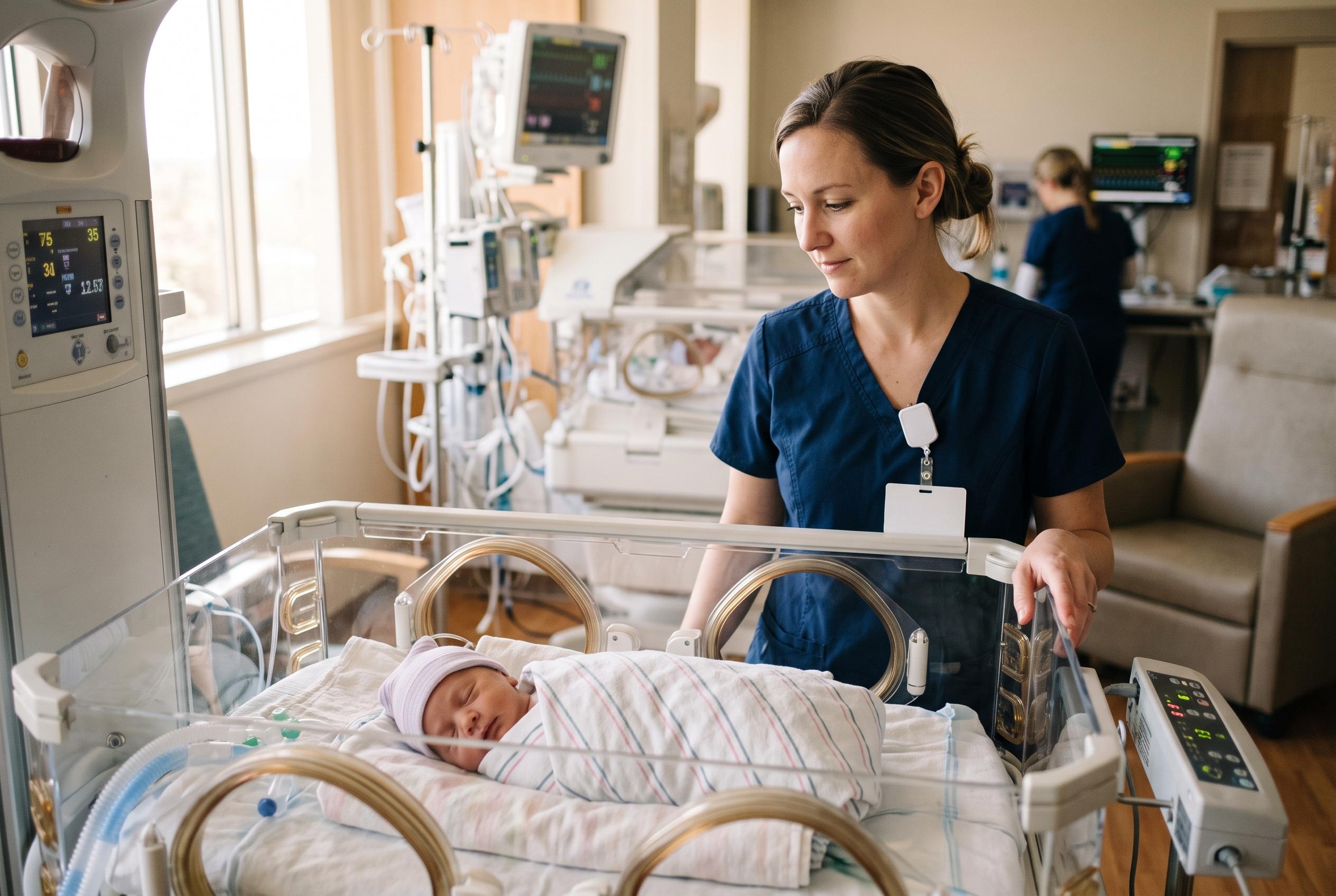

A simple measurement from routine near-term MRI scans was linked to cognitive and motor outcomes at age two in babies born as early as 22 weeks, although researchers say the finding needs wider validation.
Researchers in Vienna say a simple brain MRI measurement may help predict how extremely preterm babies develop in early childhood, offering clinicians a potentially practical way to flag risk long before standard developmental testing is possible.
The study, published in Pediatric Research, focused on infants born at the very edge of viability, between 22 and 27 weeks’ gestation. Investigators at the Medical University of Vienna analysed routine magnetic resonance scans taken near term-equivalent age, when a baby born very early reaches the stage that would have corresponded to full-term pregnancy. They then compared those images with developmental outcomes at two years of corrected age, looking for a link between the size of the brainstem and later cognitive and motor performance.
The brainstem sits at the base of the brain and controls essential functions such as breathing, heart rate, feeding coordination and other automatic reflexes. According to the Vienna team, it may also act as a marker of broader brain development in babies born far too soon. Their work builds on earlier research showing that term-equivalent MRI can help predict later outcomes in very preterm children, but it goes a step further by identifying a measurement that can be taken on a single midline image with standard software. That matters because many neonatal units already perform MRI before discharge, while more advanced imaging methods often require specialist analysis.
In the Vienna cohort, brainstem measurements increased with gestational age and were smaller in infants with severe neonatal brain injury. The researchers reported that the pons, the rounded section of the brainstem that carries major nerve pathways, was the most informative region when outcomes were analysed separately. A separate body of research has reached a similar conclusion that different MRI markers, from corpus callosum size to cortical grey matter shape and quantitative brain tissue measures, can help predict later development in preterm children, suggesting that brain growth patterns visible on scans may provide a meaningful window into neurodevelopmental risk.
The strongest findings came when the team compared brainstem size with Bayley developmental scores at two years. Higher brainstem z-scores were linked with better cognitive and motor results, even after adjustment for a range of clinical factors, including birthweight, sepsis, major intestinal disease, severe retinopathy, chronic lung disease and different forms of brain injury. The study also found that lower birthweight, higher early lactate levels and lower first-minute Apgar scores were associated with a brainstem size below the tenth percentile, pointing to the possible role of growth restriction and early physiological stress.
The authors say the measure is attractive because it is quick, low-cost and based on scans that many centres already obtain. But they also caution that the work is retrospective, from a single centre and still needs independent validation before it can be used routinely to guide prognosis. Even so, the findings add to a growing picture in neonatal imaging: small structural differences seen early in life may help identify which extremely preterm infants are most likely to need extra support, and which may do better than expected.
Disclaimer: This content is for informational purposes only and is not intended to be a substitute for professional medical judgment, advice, diagnosis, or treatment.