

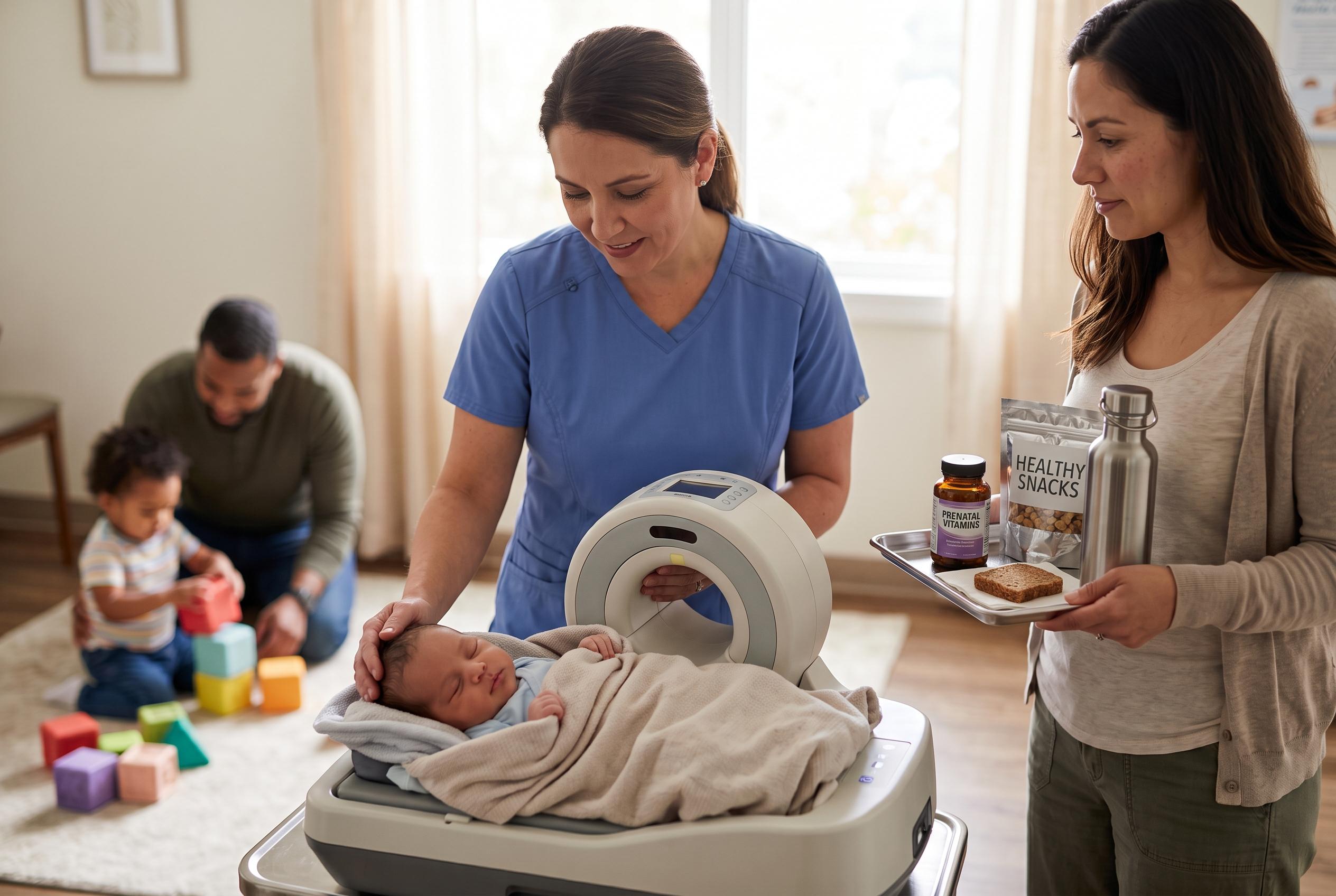

A pioneering study using portable MRI scans reveals that anaemia during pregnancy can cause measurable and widening brain volume differences in infants by age two, highlighting the importance of maternal nutrition and innovative imaging solutions in vulnerable populations.
A study from King’s College London suggests that anaemia during pregnancy may shape a baby’s brain within the first year of life, with measurable differences appearing by around 12 months and widening by age two.
Researchers followed 394 mother-infant pairs in Cape Town and used both a standard hospital MRI scanner and a portable ultra-low-field device to track brain development in a subgroup of infants between three months and two years of age. According to the study in Brain Communications, babies born to mothers who had anaemia in pregnancy had, on average, about 4% smaller total brain volume than those whose mothers were not anaemic.
The differences were seen in regions tied to movement, learning, emotion regulation, executive function and communication between the brain’s hemispheres, including the putamen, caudate nucleus and corpus callosum. The corpus callosum, which links the two sides of the brain, was about 4% smaller at 12 months in the exposed group and about 6% smaller by 24 months, suggesting the gap widened over time.
The findings add to earlier work from South Africa showing similar differences in older children, but the new research indicates they can be detected much earlier than previously documented. In a statement, Jessica Ringshaw, the study’s first author, said the results show how early these effects can be seen and underline the importance of improving treatment and prevention before and during pregnancy.
The study also points to the promise of portable MRI in places where conventional scanners are scarce. The lightweight 64 millitesla device used in the research can be taken directly into communities, needs less infrastructure than a hospital scanner and is quieter, making it easier to scan sleeping infants without sedation. Steve Williams, a professor of neuroimaging at King’s College London, said the technology could support large-scale measurement of malnutrition’s impact on the developing brain and help assess interventions earlier.
Anaemia in pregnancy affects more than a third of pregnant women worldwide, with the heaviest burden in sub-Saharan Africa and South Asia. The condition is often linked to iron deficiency, but infections such as HIV can also increase the risk. In this cohort, mothers living with HIV were more likely to be anaemic, which the researchers said may reflect inflammation interfering with iron use in the body.
Kirsten Donald, the study’s senior author, said the work opens a new way to measure how maternal nutrition affects child development in the populations most affected. Related research published elsewhere has also linked maternal and child anaemia to poorer developmental outcomes, while a systematic review has found maternal anaemia is associated with negative effects on neonatal brain development. Together, the evidence points to anaemia in pregnancy as a preventable risk factor with consequences that may begin before birth and continue through early childhood.
Disclaimer: This content is for informational purposes only and is not intended to be a substitute for professional medical judgment, advice, diagnosis, or treatment.